 Transephageal Echocardiography
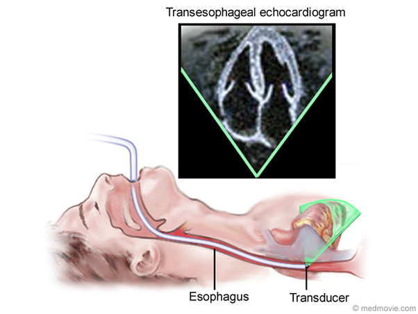
Transesophageal echocardiography (TEE) is a test that produces pictures of your heart. TEE uses high-frequency sound waves (ultrasound) to make detailed pictures of your heart and the arteries that lead to and from it. Unlike a standard echocardogram, the echo transducer that produces the sound waves for TEE is attached to a thin tube that passes through your mouth, down your throat and into your esophagus. Because the esophagus is so close to the upper chambers of the heart, very clear images of those heart structures, valves and outer lining (pericardium), as well as the blood vessels that connect to your heart can be obtained.
TEE is often used to more thoroughly evaluate for infection of the heart valves, cardiac masses or clots, abnormal communications between chambers, and diagnose aortic dissections. Patients cannot eat prior to the test but can take home medications with sips of water. The test is done in a hospital setting and the patient will need a relative or friend to drive home because it is done with sedation.